

Are Cytoskeleton In Plant And Animal Cells
| Jail cell biology | |
|---|---|
| Animate being cell diagram | |
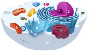 Components of a typical animal cell:
|

The cytoskeleton is a complex, dynamic network of interlinking protein filaments present in the cytoplasm of all cells, excluding leaner and archaea.[1] It extends from the jail cell nucleus to the prison cell membrane and is composed of similar proteins in the various organisms. In eukaryotes, it is equanimous of three main components, microfilaments, intermediate filaments and microtubules, and these are all capable of rapid growth or disassembly dependent on the jail cell'south requirements.[2]
A multitude of functions tin be performed by the cytoskeleton. Its principal office is to give the cell its shape and mechanical resistance to deformation, and through clan with extracellular connective tissue and other cells it stabilizes entire tissues.[3] [4] The cytoskeleton can also contract, thereby deforming the jail cell and the jail cell's surroundings and assuasive cells to migrate.[5] Moreover, it is involved in many jail cell signaling pathways and in the uptake of extracellular material (endocytosis),[6] the segregation of chromosomes during cellular segmentation,[3] the cytokinesis stage of cell partition,[vii] as scaffolding to organize the contents of the prison cell in infinite[five] and in intracellular send (for example, the motility of vesicles and organelles within the cell)[three] and tin can be a template for the construction of a jail cell wall.[iii] Furthermore, it can form specialized structures, such as flagella, cilia, lamellipodia and podosomes. The structure, function and dynamic behavior of the cytoskeleton can be very unlike, depending on organism and cell type.[3] [7] Even inside i jail cell, the cytoskeleton tin can change through clan with other proteins and the previous history of the network.[5]
A large-scale case of an activeness performed by the cytoskeleton is muscle contraction. This is carried out by groups of highly specialized cells working together. A main component in the cytoskeleton that helps show the true function of this muscle contraction is the microfilament. Microfilaments are composed of the most abundant cellular protein known equally actin.[eight] During contraction of a muscle, within each muscle cell, myosin molecular motors collectively exert forces on parallel actin filaments. Muscle contraction starts from nervus impulses which then causes increased amounts of calcium to be released from the sarcoplasmic reticulum. Increases in calcium in the cytosol allows muscle contraction to begin with the aid of 2 proteins, tropomyosin and troponin.[viii] Tropomyosin inhibits the interaction between actin and myosin, while troponin senses the increment in calcium and releases the inhibition.[9] This action contracts the muscle cell, and through the synchronous process in many muscle cells, the entire muscle.
History [edit]
In 1903, Nikolai 1000. Koltsov proposed that the shape of cells was determined by a network of tubules that he termed the cytoskeleton. The concept of a poly peptide mosaic that dynamically coordinated cytoplasmic biochemistry was proposed past Rudolph Peters in 1929[10] while the term (cytosquelette, in French) was showtime introduced past French embryologist Paul Wintrebert in 1931.[11]
When the cytoskeleton was commencement introduced, it was thought to be an uninteresting gel-like substance that helped organelles stay in identify.[12] Much research took place to try to sympathise the purpose of the cytoskeleton and its components. With the help of Stuart Hameroff and Roger Penrose, it was discovered that microtubules vibrate within neurons in the brain, suggesting that encephalon waves come from deeper microtubule vibrations.[xiii] This discovery demonstrated that the cytoskeleton is non just a gel-like substance and that information technology actually has a purpose.[ disputed ]
Initially, information technology was thought that the cytoskeleton was sectional to eukaryotes but in 1992 it was discovered to exist nowadays in prokaryotes as well. This discovery came subsequently the realization that bacteria possess proteins that are homologous to tubulin and actin; the main components of the eukaryotic cytoskeleton.[14]
Eukaryotic cytoskeleton [edit]
Eukaryotic cells contain iii chief kinds of cytoskeletal filaments: microfilaments, microtubules, and intermediate filaments. In neurons the intermediate filaments are known as neurofilaments.[xv] Each blazon is formed past the polymerization of a distinct type of protein subunit and has its own characteristic shape and intracellular distribution. Microfilaments are polymers of the protein actin and are vii nm in diameter. Microtubules are equanimous of tubulin and are 25 nm in bore. Intermediate filaments are composed of various proteins, depending on the blazon of jail cell in which they are constitute; they are normally 8-12 nm in diameter.[one] The cytoskeleton provides the cell with structure and shape, and by excluding macromolecules from some of the cytosol, it adds to the level of macromolecular crowding in this compartment.[16] Cytoskeletal elements interact extensively and intimately with cellular membranes.[17]
Enquiry into neurodegenerative disorders such every bit Parkinson's illness, Alzheimer'southward affliction, Huntington's affliction, and amyotrophic lateral sclerosis (ALS) indicate that the cytoskeleton is affected in these diseases.[18] Parkinson's disease is marked by the deposition of neurons, resulting in tremors, rigidity, and other not-motor symptoms. Research has shown that microtubule assembly and stability in the cytoskeleton is compromised causing the neurons to degrade over time.[xix] In Alzheimer's disease, tau proteins which stabilize microtubules malfunction in the progression of the illness causing pathology of the cytoskeleton.[20] Excess glutamine in the Huntington protein involved with linking vesicles onto the cytoskeleton is also proposed to be a factor in the development of Huntington's Affliction.[21] Amyotrophic Lateral Sclerosis results in a loss of movement caused by the degradation of motor neurons, and also involves defects of the cytoskeleton.[22]
Accessory proteins including motor proteins regulate and link the filaments to other jail cell compounds and each other and are essential for controlled associates of cytoskeletal filaments in particular locations.[23]
A number of minor-molecule cytoskeletal drugs have been discovered that interact with actin and microtubules. These compounds have proven useful in studying the cytoskeleton, and several take clinical applications.
Microfilaments [edit]
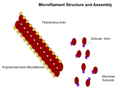
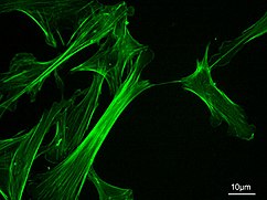
Microfilaments, as well known equally actin filaments, are equanimous of linear polymers of G-actin proteins, and generate force when the growing (plus) cease of the filament pushes against a barrier, such as the cell membrane. They also act as tracks for the movement of myosin molecules that affix to the microfilament and "walk" forth them. In general, the major component or protein of microfilaments are actin. The G-actin monomer combines to class a polymer which continues to grade the microfilament (actin filament). These subunits then assemble into ii chains that intertwine into what are called F-actin bondage.[24] Myosin motoring forth F-actin filaments generates contractile forces in so-chosen actomyosin fibers, both in musculus as well as most non-muscle cell types.[25] Actin structures are controlled by the Rho family of small GTP-binding proteins such as Rho itself for contractile acto-myosin filaments ("stress fibers"), Rac for lamellipodia and Cdc42 for filopodia.
Functions include:
- Musculus contraction
- Cell movement
- Intracellular ship/trafficking
- Maintenance of eukaryotic cell shape
- Cytokinesis
- Cytoplasmic streaming[24]
Intermediate filaments [edit]
Microscopy of keratin filaments inside cells
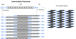
Intermediate filaments are a function of the cytoskeleton of many eukaryotic cells. These filaments, averaging 10 nanometers in diameter, are more than stable (strongly bound) than microfilaments, and heterogeneous constituents of the cytoskeleton. Like actin filaments, they function in the maintenance of cell-shape by bearing tension (microtubules, past contrast, resist pinch simply can also carry tension during mitosis and during the positioning of the centrosome). Intermediate filaments organize the internal tridimensional structure of the cell, anchoring organelles and serving as structural components of the nuclear lamina. They likewise participate in some prison cell-cell and cell-matrix junctions. Nuclear lamina be in all animals and all tissues. Some animals like the fruit wing do not have any cytoplasmic intermediate filaments. In those animals that limited cytoplasmic intermediate filaments, these are tissue specific.[4] Keratin intermediate filaments in epithelial cells provide protection for different mechanical stresses the peel may endure. They also provide protection for organs against metabolic, oxidative, and chemic stresses. Strengthening of epithelial cells with these intermediate filaments may foreclose onset of apoptosis, or jail cell death, by reducing the probability of stress.[26]
Intermediate filaments are about ordinarily known as the back up system or "scaffolding" for the cell and nucleus while also playing a office in some cell functions. In combination with proteins and desmosomes, the intermediate filaments course cell-jail cell connections and anchor the cell-matrix junctions that are used in messaging between cells also as vital functions of the prison cell. These connections allow the cell to communicate through the desmosome of multiple cells to adapt structures of the tissue based on signals from the cells environment. Mutations in the IF proteins have been shown to cause serious medical problems such as premature aging, desmin mutations compromising organs, Alexander Affliction, and muscular dystrophy.[27]
Different intermediate filaments are:
- fabricated of vimentins. Vimentin intermediate filaments are in general present in mesenchymal cells.
- made of keratin. Keratin is present in general in epithelial cells.
- neurofilaments of neural cells.
- made of lamin, giving structural support to the nuclear envelope.
- fabricated of desmin, play an important role in structural and mechanical support of muscle cells.[28]
Microtubules [edit]
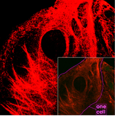
![]()
Microtubules in a gel-fixated jail cell
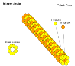
Microtubules are hollow cylinders nigh 23 nm in bore (lumen diameter of approximately 15 nm), near usually comprising 13 protofilaments that, in turn, are polymers of alpha and beta tubulin. They have a very dynamic behavior, binding GTP for polymerization. They are commonly organized past the centrosome.
In ix triplet sets (star-shaped), they form the centrioles, and in nine doublets oriented about ii boosted microtubules (cycle-shaped), they form cilia and flagella. The latter formation is unremarkably referred to as a "nine+2" organization, wherein each doublet is connected to another by the protein dynein. As both flagella and cilia are structural components of the prison cell, and are maintained by microtubules, they tin be considered part of the cytoskeleton. In that location are two types of cilia: motile and non-motile cilia. Cilia are short and more numerous than flagella. The motile cilia take a rhythmic waving or beating move compared to the non-motile cilia which receive sensory information for the prison cell; processing signals from the other cells or the fluids surrounding information technology. Additionally, the microtubules control the beating (movement) of the cilia and flagella.[29] Also, the dynein arms attached to the microtubules function as the molecular motors. The motion of the cilia and flagella is created by the microtubules sliding past ane another, which requires ATP.[29] They play key roles in:
- intracellular send (associated with dyneins and kinesins, they transport organelles like mitochondria or vesicles).
- the axoneme of cilia and flagella.

Cross section diagram through the cilium, showing the "9 + 2" arrangement of microtubules
- the mitotic spindle.
- synthesis of the jail cell wall in plants.
In addition to the roles described above, Stuart Hameroff and Roger Penrose accept proposed that microtubules function in consciousness.[xxx]
Comparison [edit]
| Cytoskeleton type[31] | Bore (nm)[32] | Structure | Subunit examples[31] |
|---|---|---|---|
| Microfilaments | six | Double helix | Actin |
| Intermediate filaments | x | Two anti-parallel helices/dimers, forming tetramers |
|
| Microtubules | 23 | Protofilaments, in plough consisting of tubulin subunits in complex with stathmin[33] | α- and β-Tubulin |
Septins [edit]
Septins are a group of the highly conserved GTP binding proteins found in eukaryotes. Different septins form poly peptide complexes with each other. These tin can assemble to filaments and rings. Therefore, septins can be considered part of the cytoskeleton.[34] The function of septins in cells include serving as a localized attachment site for other proteins, and preventing the diffusion of sure molecules from one prison cell compartment to another.[34] In yeast cells, they build scaffolding to provide structural support during prison cell division and compartmentalize parts of the jail cell. Recent research in homo cells suggests that septins build cages around bacterial pathogens, immobilizing the harmful microbes and preventing them from invading other cells.[35]
Spectrin [edit]
Spectrin is a cytoskeletal protein that lines the intracellular side of the plasma membrane in eukaryotic cells. Spectrin forms pentagonal or hexagonal arrangements, forming a scaffolding and playing an important part in maintenance of plasma membrane integrity and cytoskeletal structure.[36]
Yeast cytoskeleton [edit]
In budding yeast (an important model organism), actin forms cortical patches, actin cables, and a cytokinetic ring and the cap. Cortical patches are detached actin bodies on the membrane and are vital for endocytosis, especially the recycling of glucan synthase which is important for cell wall synthesis. Actin cables are bundles of actin filaments and are involved in the ship of vesicles towards the cap (which contains a number of dissimilar proteins to polarize prison cell growth) and in the positioning of mitochondria. The cytokinetic ring forms and constricts around the site of cell partitioning.[37]
Prokaryotic cytoskeleton [edit]
Prior to the work of Jones et al., 2001, the prison cell wall was believed to be the deciding factor for many bacterial cell shapes, including rods and spirals. When studied, many misshapen bacteria were establish to have mutations linked to development of a cell envelope.[38] The cytoskeleton was in one case thought to be a feature only of eukaryotic cells, merely homologues to all the major proteins of the eukaryotic cytoskeleton accept been found in prokaryotes.[39] Harold Erickson notes that earlier 1992, just eukaryotes were believed to have cytoskeleton components. Still, enquiry in the early '90s suggested that bacteria and archaea had homologues of actin and tubulin, and that these were the basis of eukaryotic microtubules and microfilaments.[xl] Although the evolutionary relationships are and so afar that they are not obvious from poly peptide sequence comparisons alone, the similarity of their three-dimensional structures and similar functions in maintaining cell shape and polarity provides strong evidence that the eukaryotic and prokaryotic cytoskeletons are truly homologous.[41] Three laboratories independently discovered that FtsZ, a protein already known every bit a primal player in bacterial cytokinesis, had the "tubulin signature sequence" present in all α-, β-, and γ-tubulins.[twoscore] Nonetheless, some structures in the bacterial cytoskeleton may not have been identified as of yet.[25] [42]
FtsZ [edit]
FtsZ was the outset poly peptide of the prokaryotic cytoskeleton to be identified. Like tubulin, FtsZ forms filaments in the presence of guanosine triphosphate (GTP), only these filaments practise not grouping into tubules. During cell partition, FtsZ is the first protein to move to the segmentation site, and is essential for recruiting other proteins that synthesize the new jail cell wall betwixt the dividing cells.
MreB and ParM [edit]
Prokaryotic actin-similar proteins, such as MreB, are involved in the maintenance of cell shape. All not-spherical bacteria have genes encoding actin-like proteins, and these proteins form a helical network beneath the cell membrane that guides the proteins involved in cell wall biosynthesis.[43]
Some plasmids encode a carve up arrangement that involves an actin-like poly peptide ParM. Filaments of ParM exhibit dynamic instability, and may sectionalization plasmid DNA into the dividing girl cells by a machinery analogous to that used by microtubules during eukaryotic mitosis.[25] [44]
Crescentin [edit]
The bacterium Caulobacter crescentus contains a third protein, crescentin, that is related to the intermediate filaments of eukaryotic cells. Crescentin is also involved in maintaining cell shape, such as helical and vibrioid forms of bacteria, but the mechanism by which it does this is currently unclear.[45] Additionally, curvature could exist described by the displacement of crescentic filaments, after the disruption of peptidoglycan synthesis.[46]
Mutual features and differences between prokaryotes and eukaryotes [edit]
By definition, the cytoskeleton is composed of proteins that can class longitudinal arrays (fibres) in all organisms. These filament forming proteins accept been classified into four classes. Tubulin-like, actin-like, Walker A cytoskeletal ATPases (WACA-proteins), and intermediate filaments.[7] [25]
Tubulin-similar proteins are tubulin in eukaryotes and FtsZ, TubZ, RepX in prokaryotes. Actin-like proteins are actin in eukaryotes and MreB, FtsA in prokaryotes. An instance of a WACA-proteins, which are generally institute in prokaryotes, is MinD. Examples for intermediate filaments, which have almost exclusively been found in animals (i.e. eukaryotes) are the lamins, keratins, vimentin, neurofilaments, and desmin.[vii]
Although tubulin-similar proteins share some amino acid sequence similarity, their equivalence in protein-fold and the similarity in the GTP binding site is more hitting. The same holds true for the actin-similar proteins and their structure and ATP binding domain.[7] [25]
Cytoskeletal proteins are ordinarily correlated with cell shape, DNA segregation and cell division in prokaryotes and eukaryotes. Which proteins fulfill which task is very different. For example, Dna segregation in all eukaryotes happens through use of tubulin, only in prokaryotes either WACA proteins, actin-like or tubulin-similar proteins tin can be used. Jail cell segmentation is mediated in eukaryotes past actin, but in prokaryotes usually past tubulin-like (frequently FtsZ-ring) proteins and sometimes (Crenarchaeota) ESCRT-Iii, which in eukaryotes still has a office in the final step of division.[7]
Cytoplasmic streaming [edit]
Move of organelles in Tradescantia stamen hair cells
Cytoplasmic streaming, also known every bit cyclosis, is the agile movement of a jail cell's contents along the components of the cytoskeleton. While mainly seen in plants, all cell types use this process for transportation of waste, nutrients, and organelles to other parts of the jail cell.[47] Constitute and algae cells are generally larger than many other cells; so cytoplasmic streaming is important in these types of cells. This is considering the cell'southward extra book requires cytoplasmic streaming in order to move organelles throughout the entire prison cell.[48] Organelles move along microfilaments in the cytoskeleton driven by myosin motors binding and pushing forth actin filament bundles.[47]
See likewise [edit]
- Nuclear matrix – Fibrillar network lying on nuclear membrane
- Cell cortex – Layer on the inner face up of a prison cell membrane
References [edit]
- ^ a b Hardin J, Bertoni K, Kleinsmith LJ (2015). Becker'due south World of the Cell (eighth ed.). New York: Pearson. pp. 422–446. ISBN978013399939-6.
- ^ McKinley, Michael; Dean O'Loughlin, Valerie; Pennefather-O'Brien, Elizabeth; Harris, Ronald (2015). Human Beefcake (4th ed.). New York: McGraw Hill Instruction. p. 29. ISBN978-0-07-352573-0.
- ^ a b c d east Alberts B, et al. (2008). Molecular Biology of the Cell (fifth ed.). New York: Garland Science. ISBN978-0-8153-4105-v.
- ^ a b Herrmann H, Bär H, Kreplak L, Strelkov SV, Aebi U (July 2007). "Intermediate filaments: from cell architecture to nanomechanics". Nature Reviews. Molecular Cell Biology. 8 (seven): 562–73. doi:x.1038/nrm2197. PMID 17551517. S2CID 27115011.
- ^ a b c Fletcher DA, Mullins RD (January 2010). "Cell mechanics and the cytoskeleton". Nature. 463 (7280): 485–92. Bibcode:2010Natur.463..485F. doi:10.1038/nature08908. PMC2851742. PMID 20110992.
- ^ Geli MI, Riezman H (April 1998). "Endocytic internalization in yeast and creature cells: similar and unlike". Journal of Jail cell Science. 111 ( Pt 8) (8): 1031–7. doi:10.1242/jcs.111.8.1031. PMID 9512499.
- ^ a b c d e f Wickstead B, Gull Thousand (August 2011). "The development of the cytoskeleton". The Periodical of Jail cell Biology. 194 (4): 513–25. doi:10.1083/jcb.201102065. PMC3160578. PMID 21859859.
- ^ a b Cooper, Geoffrey M. (2000). "Actin, Myosin, and Prison cell Motion". The Jail cell: A Molecular Arroyo. 2d Edition. Archived from the original on 2018-04-28.
- ^ Berg JM, Tymoczko JL, Stryer Fifty (2002). "Myosins Move Along Actin Filaments". Biochemistry. 5th Edition. Archived from the original on 2018-05-02.
- ^ Peters RA. "The Harben Lectures, 1929. Reprinted in: Peters, R. A. (1963) Biochemical lesions and lethal synthesis, p. 216. Pergamon Press, Oxford".
- ^ Frixione E (June 2000). "Recurring views on the construction and function of the cytoskeleton: a 300-year ballsy". Cell Motility and the Cytoskeleton. 46 (2): 73–94. doi:ten.1002/1097-0169(200006)46:2<73::Aid-CM1>three.0.CO;ii-0. PMID 10891854. S2CID 16728876.
- ^ Hardin J (2015-12-03). Becker's World of the Cell (9th ed.). Pearson. p. 351. ISBN978-0-321-93492-v.
- ^ Elsevier. "Discovery of Quantum Vibrations in "Microtubules" Inside Brain Neurons Corroborates Controversial 20-Year-Old Theory of Consciousness". www.elsevier.com. Archived from the original on 2016-eleven-07. Retrieved 2017-11-20 .
- ^ Wickstead B, Dupe K (August 2011). "The evolution of the cytoskeleton". The Journal of Cell Biology. 194 (four): 513–25. doi:ten.1083/jcb.201102065. PMC3160578. PMID 21859859.
- ^ Taran, AS; Shuvalova, LD; Lagarkova, MA; Alieva, IB (22 June 2020). "Huntington's Disease-An Outlook on the Coaction of the HTT Protein, Microtubules and Actin Cytoskeletal Components". Cells. ix (6): 1514. doi:10.3390/cells9061514. PMC7348758. PMID 32580314.
- ^ Minton AP (Oct 1992). "Confinement as a determinant of macromolecular construction and reactivity". Biophysical Journal. 63 (4): 1090–100. Bibcode:1992BpJ....63.1090M. doi:10.1016/S0006-3495(92)81663-6. PMC1262248. PMID 1420928.
- ^ Doherty GJ, McMahon HT (2008). "Arbitration, modulation, and consequences of membrane-cytoskeleton interactions". Annual Review of Biophysics. 37: 65–95. doi:ten.1146/annurev.biophys.37.032807.125912. PMID 18573073. S2CID 17352662.
- ^ Pelucchi, Silvia; Stringhi, Ramona; Marcello, Elena (2020). "Dendritic Spines in Alzheimer's Disease: How the Actin Cytoskeleton Contributes to Synaptic Failure". International Journal of Molecular Sciences. 21 (3): 908. doi:10.3390/ijms21030908. ISSN 1422-0067. PMC7036943. PMID 32019166.
- ^ Pellegrini L, Wetzel A, Grannó S, Heaton G, Harvey K (February 2017). "Back to the tubule: microtubule dynamics in Parkinson'south disease". Cellular and Molecular Life Sciences. 74 (3): 409–434. doi:10.1007/s00018-016-2351-6. PMC5241350. PMID 27600680.
- ^ Bamburg JR, Bloom GS (August 2009). "Cytoskeletal pathologies of Alzheimer's Disease". Cell Motion and the Cytoskeleton. 66 (eight): 635–49. doi:10.1002/cm.20388. PMC2754410. PMID 19479823.
- ^ Caviston JP, Holzbaur EL (April 2009). "Huntingtin protein is an essential integrator of intracellular vesicular trafficking". Trends in Cell Biology. nineteen (iv): 147–55. doi:ten.1016/j.tcb.2009.01.005. PMC2930405. PMID 19269181.
- ^ Julien JP, Millecamps S, Kriz J (2005). "Cytoskeletal Defects in Amyotrophic Lateral Sclerosis (motor neuron affliction)". Novartis Foundation Symposium. 264: 183–92, give-and-take 192–6, 227–30. PMID 15773754.
- ^ Alberts, Bruce (2015). Molecular Biology of the Jail cell. Garland Science. p. 889. ISBN978-0-8153-4464-3.
- ^ a b Cooper, Geoffrey G. (2000). "Construction and Organization of Actin Filaments". The Cell: A Molecular Approach. 2nd Edition. Archived from the original on 2018-05-02.
- ^ a b c d e Gunning Prisoner of war, Ghoshdastider U, Whitaker Southward, Popp D, Robinson RC (June 2015). "The evolution of compositionally and functionally singled-out actin filaments". Journal of Cell Science. 128 (11): 2009–19. doi:ten.1242/jcs.165563. PMID 25788699.
- ^ Pan X, Hobbs RP, Coulombe PA (February 2013). "The expanding significance of keratin intermediate filaments in normal and diseased epithelia". Current Opinion in Prison cell Biology. 25 (1): 47–56. doi:10.1016/j.ceb.2012.10.018. PMC3578078. PMID 23270662.
- ^ Herrmann H, Bär H, Kreplak L, Strelkov SV, Aebi U (July 2007). "Intermediate filaments: from cell architecture to nanomechanics". Nature Reviews. Molecular Cell Biology. 8 (7): 562–73. doi:10.1038/nrm2197. PMID 17551517. S2CID 27115011.
- ^ Paulin D, Li Z (November 2004). "Desmin: a major intermediate filament protein essential for the structural integrity and function of muscle". Experimental Cell Inquiry. 301 (1): 1–seven. doi:10.1016/j.yexcr.2004.08.004. PMID 15501438.
- ^ a b Lodish, Harvey; Berk, Arnold; Zipursky, S. Lawrence; Matsudaira, Paul; Baltimore, David; Darnell, James (2 May 2018). "Cilia and Flagella: Structure and Motion". Archived from the original on 2 May 2018. Retrieved 2 May 2018 – via world wide web.ncbi.nlm.nih.gov.
- ^ Hameroff, S. and Penrose, R. Physics of Life Reviews 2014, 11, 39-78
- ^ a b Unless else specified in boxes, then ref is:Boron WF (2003). Medical Physiology: A Cellular And Molecular Approaoch. Elsevier/Saunders. p. 1300. ISBN978-1-4160-2328-9. Page 25
- ^ Fuchs Due east, Cleveland DW (January 1998). "A structural scaffolding of intermediate filaments in health and disease". Science. 279 (5350): 514–9. Bibcode:1998Sci...279..514F. doi:10.1126/science.279.5350.514. PMID 9438837.
- ^ Steinmetz MO (May 2007). "Structure and thermodynamics of the tubulin-stathmin interaction". Journal of Structural Biology. 158 (2): 137–47. doi:x.1016/j.jsb.2006.07.018. PMID 17029844.
- ^ a b Mostowy Southward, Cossart P (February 2012). "Septins: the quaternary component of the cytoskeleton". Nature Reviews. Molecular Cell Biological science. 13 (three): 183–94. doi:10.1038/nrm3284. PMID 22314400. S2CID 2418522.
- ^ Mascarelli A (December 2011). "Septin proteins take bacterial prisoners: A cellular defense confronting microbial pathogens holds therapeutic potential". Nature. doi:10.1038/nature.2011.9540. S2CID 85080734.
- ^ Huh GY, Glantz SB, Je Due south, Morrow JS, Kim JH (Dec 2001). "Calpain proteolysis of alpha II-spectrin in the normal developed human brain". Neuroscience Letters. 316 (i): 41–4. doi:10.1016/S0304-3940(01)02371-0. PMID 11720774. S2CID 53270680.
- ^ Pruyne D, Bretscher A (February 2000). "Polarization of cell growth in yeast". Journal of Cell Science. 113 ( Pt 4) (iv): 571–85. doi:ten.1242/jcs.113.iv.571. PMID 10652251.
- ^ Jones, Laura J. F.; Carballido-López, Rut; Errington, Jeffery (2001-03-23). "Control of Cell Shape in Bacteria: Helical, Actin-similar Filaments in Bacillus subtilis". Cell. 104 (half dozen): 913–922. doi:ten.1016/S0092-8674(01)00287-2. PMID 11290328. S2CID 14207533.
- ^ Shih YL, Rothfield L (September 2006). "The bacterial cytoskeleton". Microbiology and Molecular Biology Reviews. 70 (3): 729–54. doi:10.1128/MMBR.00017-06. PMC1594594. PMID 16959967.
- ^ a b Erickson HP (February 2017). "The discovery of the prokaryotic cytoskeleton: 25th anniversary". Molecular Biology of the Cell. 28 (three): 357–358. doi:10.1091/mbc.E16-03-0183. PMC5341718. PMID 28137947.
- ^ Michie KA, Löwe J (2006). "Dynamic filaments of the bacterial cytoskeleton" (PDF). Almanac Review of Biochemistry. 75: 467–92. doi:ten.1146/annurev.biochem.75.103004.142452. PMID 16756499.
- ^ Briegel A, Dias DP, Li Z, Jensen RB, Frangakis AS, Jensen GJ (October 2006). "Multiple large filament bundles observed in Caulobacter crescentus by electron cryotomography". Molecular Microbiology. 62 (i): 5–14. doi:ten.1111/j.1365-2958.2006.05355.x. PMID 16987173.
- ^ Popp D, Narita A, Maeda K, Fujisawa T, Ghoshdastider U, Iwasa M, Maéda Y, Robinson RC (May 2010). "Filament structure, organization, and dynamics in MreB sheets". The Periodical of Biological Chemistry. 285 (21): 15858–65. doi:10.1074/jbc.M109.095901. PMC2871453. PMID 20223832.
- ^ Popp D, Narita A, Lee LJ, Ghoshdastider U, Xue B, Srinivasan R, Balasubramanian MK, Tanaka T, Robinson RC (June 2012). "Novel actin-like filament structure from Clostridium tetani". The Periodical of Biological Chemistry. 287 (25): 21121–9. doi:10.1074/jbc.M112.341016. PMC3375535. PMID 22514279.
- ^ Ausmees Due north, Kuhn JR, Jacobs-Wagner C (December 2003). "The bacterial cytoskeleton: an intermediate filament-like function in cell shape". Prison cell. 115 (6): 705–13. doi:x.1016/S0092-8674(03)00935-eight. PMID 14675535. S2CID 14459851.
- ^ Esue, Osigwe (January 2010). "Dynamics of the Bacterial Intermediate Filament Crescentin In Vitro and In Vivo". PLOS I. 5 (1): e8855. Bibcode:2010PLoSO...five.8855E. doi:10.1371/journal.pone.0008855. PMC2816638. PMID 20140233. Retrieved 12 September 2017.
- ^ a b Woodhouse FG, Goldstein RE (August 2013). "Cytoplasmic streaming in found cells emerges naturally by microfilament self-organization". Proceedings of the National University of Sciences of the U.s. of America. 110 (35): 14132–7. arXiv:1308.6422. Bibcode:2013PNAS..11014132W. doi:10.1073/pnas.1302736110. PMC3761564. PMID 23940314.
- ^ Goldstein RE, van de Meent JW (August 2015). "A physical perspective on cytoplasmic streaming". Interface Focus. five (four): 20150030. doi:10.1098/rsfs.2015.0030. PMC4590424. PMID 26464789.
External links [edit]
- Cytoskeleton Monthly News and Blog
- MBInfo - Cytoskeleton Dynamics
- Cytoskeleton, Cell Motility and Motors - The Virtual Library of Biochemistry, Molecular Biology and Cell Biology
- Cytoskeleton database, clinical trials, recent literature, lab registry ...
- Animation of leukocyte adhesion (Animation with some images of actin and microtubule associates and dynamics.)
- http://cellix.imba.oeaw.ac.at/ Cytoskeleton and cell motility including videos
- Open up access review commodity on the emergent complexity of the cytoskeleton (appeared in Advances in Physics, 2013)
Source: https://en.wikipedia.org/wiki/Cytoskeleton
Posted by: alvarezralmy1981.blogspot.com
0 Response to "Are Cytoskeleton In Plant And Animal Cells"
Post a Comment